Products / Services
  • Products / Services
  • Companies
  • Buy Leads
Post Buy Requirement

Zeiss Xradia 810 Ultra 3D X-Ray Microscope

Listing ID #3616812

  • Supply Type Supplier
  • Preferred Buyer Location India ( Delhi )

We are here to guide our clients with the secure payment procedures. With us, the customers get convenience to pay through both online and offline payment methods We use premium-packaging materials....
View More Details
Send Enquiry

Company Information

  • Member Since 8 Years
  • Nature of Business Retailer

Ask for more detail from the seller

Contact Supplier

Product Details no_img_icon

We are here to guide our clients with the secure payment procedures. With us, the customers get convenience to pay through both online and offline payment methods We use premium-packaging materials to safeguard our products from mechanical and environmental conditions. For our patrons, we offer customized packaging solutions as well.


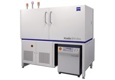
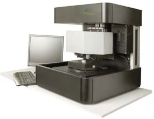
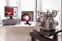
Explore at the speed of science : Achieve spatial resolution down to 50 nm with ZEISS Xradia 810 Ultra X-ray microscope, the highest among lab-based X-ray imaging systems. Experience unparalleled performance and flexibility with the non-destructive 3D imaging that plays a vital role in today’s breakthrough research. The innovative Xradia Ultra architecture, with unique X-ray optics adapted from synchrotron technology, features absorption and phase contrast. Now with energy at 5.4 keV you can increase the throughput of your nanoscale imaging by up to a factor of 10. Achieve even better contrast and image quality for medium to low Z samples with the lower energy of Xradia 810 Ultra. Expect to accomplish advanced in situ and 4D capabilities for studying structural evolution over time and under varying conditions. Extend the limits of exploration with 3D X-ray imaging for materials research, life sciences, natural resources, and diverse industrial applications.
Highest resolution, even higher contrast, faster : ZEISS solutions deliver the world’s only non-destructive 3D X-ray imaging with resolution down to 50 nm in a laboratory instrument. Along with both absorption and Zernike phase contrast, ZEISS Xradia 810 Ultra employs advanced optics adapted from the synchrotron to deliver industry-best resolution and contrast for your research. This innovative instrument enables breakthrough research by adding a critical, non-destructive step to your traditional imaging workflow.
By delivering higher contrast for your studies at 5.4 keV, Xradia 810 Ultra makes high-resolution X-ray imaging viable for a variety of difficult-to-image materials. Optimize your imaging with absorption and phase contrast for a diverse range of materials such as polymers, oxides, composites, fuel cells, geological samples and biological materials. Having pioneered nanoscale X-ray imaging at synchrotrons and prominent lab facilities worldwide, ZEISS XRM deliver ground¬breaking solutions to help put your studies at the forefront of research.
By making nanoscale X-ray imaging an order of magnitude faster, Xradia 810 Ultra optimizes the business case for XRM, whether your work is for science or industry. For central microscopy labs, a faster workflow translates into the ability to allow more users to leverage the instrument in less time, which in turn extends XRM to a broader base of subscribers. Similarly, you can quickly perform and repeat 4D and in situ studies of internal structures, making these techniques viable for many more applications. And if your applications are very targeted, such as digital rock physics used to explore feasibility of oil and gas extraction, Xradia 810 Ultra delivers measurements you can use to characterize critical parameters such as porosity within a matter of hours.


Benefits :

  • The highest resolution—down to 50 nanometers—3D X-ray imaging available in a laboratory
  • Non-destructive 3D X-ray imaging allows repeated imaging of the same sample to provide you with direct observation of microstructural evolution
  • Both absorption and Zernike phase contrast to image a diverse range of materials—medium to low Z, carbonates to shale, tissue to biomechanisms—faster by a factor of 10 at the nanoscale
  • Synchrotron-like results in your lab without the researcher’s challenge of limited access to synchrotrons, or for making your synchrotron time more efficient
  • Improved economics based on faster image acquisition times to expand the reach of the central microscopy lab to a wider range of researchers
  • Switchable field of view from 16 to 65 µm, as best suited to your imaging needs
  • Maintain high resolution while imaging of samples within in situ devices
  • Automated image alignment for tomographic reconstruction
  • Develop, prepare and test your planned synchrotron experiments in your laboratory to make limited availability of synchrotron beam time more efficient
  • Now with Scout-and-Scan Control System with an easy workflow-based user interface, ideal for the central imaging lab where users may have a wide variety of experience levels


Applications

  • Materials Research - Optimize study and design of functional materials: batteries, fuel cells, catalysts, composites, construction materials. Obtain realistic 3D microstructure data to improve computational models for bottom-up design of materials. Study and predict material properties and nanostructural evolution. Examine materials for porosity, cracks, and phase distribution in hours rather than days. Use 3D mapping for deeper understanding of properties and behaviors: porosity/pore connectivity, fiber orientation, crack propagation, particle size/distribution, delamination. High resolution, non-destructive imaging facilitates 4D and in situ studies, with high contrast for medium to low Z materials.
  • Raw Materials - Use nanoscale X-ray microscopy to determine structure of unconventional reservoir rock (carbonate, shale), obtaining parameters for characterization within hours (porosity, permeability) used in flow simulations to optimize extraction. Achieve nanoscale pore structure measurements faster by a factor of 10 for digital rock physics and special core analysis, significantly reducing time to results. Understand geomechanics under load, study the effects of tensile pressure on metals, or analyze ceramics under pressure.
  • Life Sciences - Image soft and hard tissue: microtubules in dentin, osteocyte lacunae and canaliculi in bone, bioscaffolds for tissue engineering, nanoparticle agglomerations in organic materials.
  • Electronics - Optimize your package development process through nanoscale visualization of semiconductor samples for electronics packaging research and development.


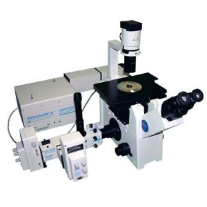
Understand nanostructural changes in 3D under load : ZEISS Xradia Ultra Load Stage uniquely enables in situ nanomechanical testing - compression, tension, indentation - with non-destructive 3D imaging. Study the evolution of interior structures in 3D, under load, down to 50 nm resolution. Understand how deformation events and failure relate to local nanoscale features. Complement existing mechanical testing methods to gain insight into behavior across multiple length scales.


Highlights

  • Add in situ nanomechanical testing capabilities to your Xradia Ultra nanoscale 3D X-ray microscope (XRM)
  • Acquire 3D tomograms of your sample under load with resolution down to 50 nm
  • Perform a variety of nanomechanical tests such as compression, tension, and indentation
  • Study a wide range of materials including metals, ceramics, composites, polymers and biomaterials
  • Complement your mechanical test results from electron microscopy, microCT and stand-alone test set-ups to understand behavior across multiple length scales: from the atomic level and the nanoscale to the micro and macro scale.
  • Available in two models with different force measurement:
    • LS108: 0.8 N maximum force
    • LS190: 9 N maximum force
  • Compatible with:
    • ZEISS Xradia 800 Ultra
    • ZEISS Xradia 810 Ultra
    • Xradia UltraXRM-L200
    • Xradia nanoXCT-200


How it works : ZEISS Xradia Ultra Load Stage can be easily configured by the user. It comprises a piezomechanical actuator with closed loop position control, a strain gauge force sensor and sets of top and bottom anvils that enable the various modes. The sample is mounted between two anvils and a sensor measures the force on the sample as a function of anvil displacement.


Key applications :  In situ nanomechanical testing is relevant for a broad range of applications covering both engineered and natural materials.
Examples include:

  • High strength alloys
  • Biomaterials / biomechanics
  • Coatings
  • Building materials
  • Fibers / composites
  • Foams


Tell us your Buy Requirement to Get Instant Response
Tell us what you need?

By clicking Get Quotes Now, I accept the T&C and Privacy Policy.

Looking for Zeiss Xradia 810 Ultra 3D X-Ray Microscope?

Quantity
Seller Contact Details
Waiting for permission
To search by voice, go to your browser settings and allow access to microphone

Allow microphone access to search with voice