Listing ID #3616809
Company Information
Ask for more detail from the seller
Contact SupplierBy using quality packing material, we make sure that the products are properly packed and there will be zero damage during transit. Moreover, our packaging experts stringently check the entire lot before making the final dispatch. We have streamlined inventory system and spacious warehouse that assist us in meeting the voluminous requirements of the clients and that too in the stipulated time span. With the latest facilities, we efficiently ensure year round availability of the products.
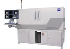
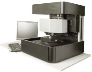
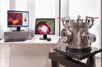
Bridge the Gap in Lab-Based Non-Destructive Submicron Microscopy : Xradia 410 Versa bridges the gap between high-performing X-ray microscopes and less powerful computed tomography (CT) systems. Delivering non-destructive 3D imaging with industry best resolution, contrast, and in situ capabilities, Xradia 410 Versa enables you to achieve groundbreaking research for the widest range of sample sizes. Enhance imaging workflows with this powerful, cost-efficient "workhorse" solution, even in diverse lab environments.
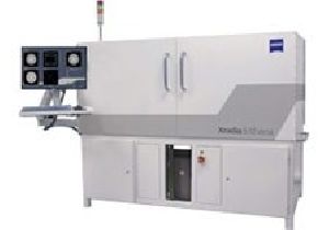
Industry-leading 4D and In Situ Capabilities for Flexible Sample Sizes and Types : Xradia 410 Versa X-ray microscope delivers cost-efficient, flexible 3D imaging to enable you to address a wide range of samples and research environments. Non-destructive X-ray imaging preserves and extends the use of your valuable samples over time. The instrument achieves 0.9 μm true spatial resolution with minimum achievable voxel size of 100 nm. Advanced absorption and phase contrast (for soft or low-Z materials) provide you with more versatility to overcome the limitations of traditional computed tomography (CT) approaches.
Xradia Versa solutions extend scientific research beyond the limits of projection-based micro- and nano-CT systems. Where traditional tomography relies on a single stage of geometric magnification, Xradia 410 Versa features a unique two-stage process based on synchrotron-caliber optics. It is easy to use, with flexible contrast. Breakthrough Resolution at a Distance (RaaD) enables you to maintain submicron resolution across a broad spectrum of sample dimensions in native environments and within a wide range of in situ rigs. Non-destructive multi-length scale capabilities allow you to image the same sample across a wide range of magnifications, making it possible to uniquely characterize the evolution of material microstructure properties between sequential treatments (4D) or as they are subjected to simulated environmental conditions (in situ).
Additionally, the Scout-and-Scan control system enables an efficient workflow environment with recipe-based set-up that makes Xradia 410 Versa easy for users with a wide variety of experience levels.
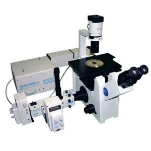
Benefits : Xradia Versa architecture uses a two-stage magnification technique to enable you to uniquely achieve resolution at a distance (RaaD). Enlarge sample images through geometric magnification as with conventional micro-CT. In the second stage, a scintillator converts X-rays to visible light, which is then optically magnified. Reducing dependence upon geometric magnification enables Xradia Versa instruments to maintain submicron resolution at large working distances. This enables you to study the widest range of sample sizes effectively, including within in situ chambers.
Applications
Use Our Super Simple Control System to Create Efficient Workflows : All of the features on the Xradia 410 Versa are seamlessly integrated within the Scout-and-Scan Control System, an efficient workflow environment that allows you to easily scout a region of interest and specify scanning parameters. The easy-to-use system is ideal for a central lab-type setting where your users may have a wide variety of experience levels. The interface maintains the flexibility for which Xradia Versa systems are known, enabling you to set-up scans even more easily. Scout-and-Scan software also offers recipe-based repeatability, which is especially useful for your in situ and 4D research, and enables you to have greater control and efficiency for future work.
New on Version 11:
Visualization and Analysis Software : ZEISS recommends Dragonfly Pro from Object Research Systems (ORS). An advanced analysis and visualization software solution for your 3D data acquired by a variety of technologies including X-ray, FIB-SEM, SEM and helium ion microscopy.
Formerly Visual SI Advanced, Dragonfly Pro delivers high-definition visualization techniques and industry-leading graphics. Dragonfly Pro supports customization through easy to use Python scripting. Users now have total control of their 3D data post-processing environment and workflows.


Connect with us