Company Information
Ask for more detail from the seller
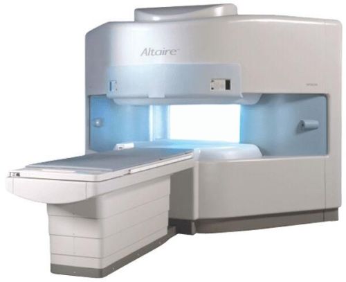
Contact SupplierThe Hitachi Altaire .7 Tesla Open MRI offers high-field performance and the patient comfort inherent in open MR systems.The Altaire .7T boasts Hitachi’s VOSI technology, optimizing each sub-system’s performance with the other sub-systems.
Altaire’s superconducting 0.7T magnet and powerful subsystems provide many of the advanced capabilities found on lower tier 1.5T systems including parallel imaging, DWI, whole body RF fat sat, time resolved MRA and more.
This performance is coupled with Hitachi’s unique gantry design and large vertical opening, improved patient comfort and expanded clinical utility to Open MRI.
Maximum advantage of coil technology, vertical field magnet and rapid gradient system.
Altaire High-Field Open MRI offers the best imaging because of its superior contrast, differentiation between muscle, fat, vessels, tendons, ligament, cartilage, cortical bone, and marrow bone space.
Ultra-wide open gantry is designed for ideal demanding cases such as anxious, claustrophobic and larger patient and reduces patient stress.
Superior image quality and diagnostic performance to all other open MRI systems. Also enhances patient comfort and facilitates ease of scanning.
50% faster patient study times than other MRI system with advanced technology for superb image resolution.
Patient friendly, wide-open design offers a panoramic view out all four sides to create a relaxing environment.
CLINICAL APPLICATION Whole body
CONFIGURATION Vertical Field, Open MRI
SURFACE COILS DualQuad T/R Body Coil, MA Head, MA C-Spine, MA Shoulder, MA Wrist, MA CTL Spine, MA Knee, MA TMJ, MA Flex Body (3 sizes), Neck, small and large Extremity, PVA (WIP), Breast (WIP), Neurovascular (WIP), Cardiac (WIP) and MA Foot//Ankle (WIP)
SPECTROSCOPY No
SYNCHRONIZATION Cardiac gating, ECG/peripheral, respiratory gating (2 modes)
PULSE SEQUENCES SE, GE, GR, IR, FIR, STIR, ss-FSE, FSE, DE-FSE/FIR, FLAIR, ss/ms-EPI, ss/ms EPI- DWI, SSP, MTC, SE/GE-EPI, MRCP, SARGE, RSSG, TRSG, BASG, Angiography: CE, PC, 2D/3D TOF
IMAGING MODES Single, multislice, volume study
TR SE: 30 – 10,000msec GE: 3.6 – 10,000msec IR: 50 – 16,700msec FSE: 200 – 16,7000msec
TE SE : 8 – 250msec IR: 5.2 -7,680msec GE: 1.8 – 2,000 msec FSE: 5.2 – 7,680
SINGLE/MULTI SLICE 0.05 sec/image (256 x 256)
FOV 5cm to 45 cm continuous
SLICE THICKNESS 2D: 2 – 100 mm; 3D: 0.5 – 5 mm
DISPLAY MATRIX 1280 x 1024
MEASURING MATRIX 512 x 512
PIXEL INTENSITY Level Range: -2,000 to +4,000
SPATIAL RESOLUTION Sub millimeter
MAGNET TYPE Self-shielded, superconducting

Connect with us